CD115 (c-fms) Monoclonal Antibody (12-3A3-1B10), PE-Cyanine7, eBioscience
PRODUCT DETAILS
Host: Rat
Isotype: IgG2a, kappa
Clonality: Monoclonal
Clone: 12-3A3-1B10
Format: PE-Cyanine7
Reactivity: Hu
Application: Flow Cytometry
Tested Dilution: 5 µL (0.03 µg)/test
Concentration: 5 μL/Test
Storage: 4°C, store in dark, DO NOT FREEZE!
Formulation: PBS with BSA and 0.09% sodium azide; pH 7.2
Purification: Affinity chromatography
Data Sheet: TDS
Specific Information
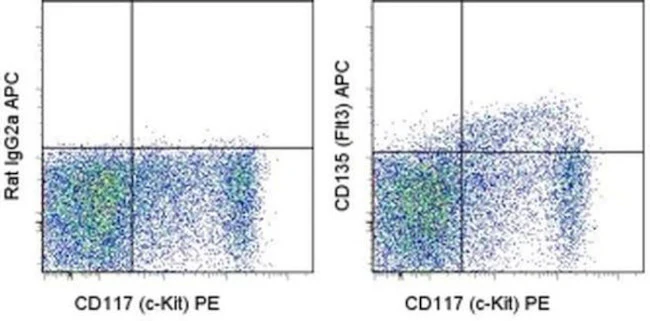
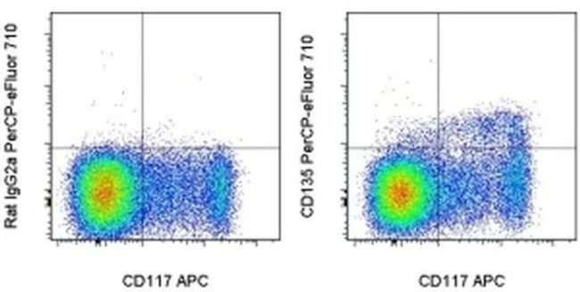
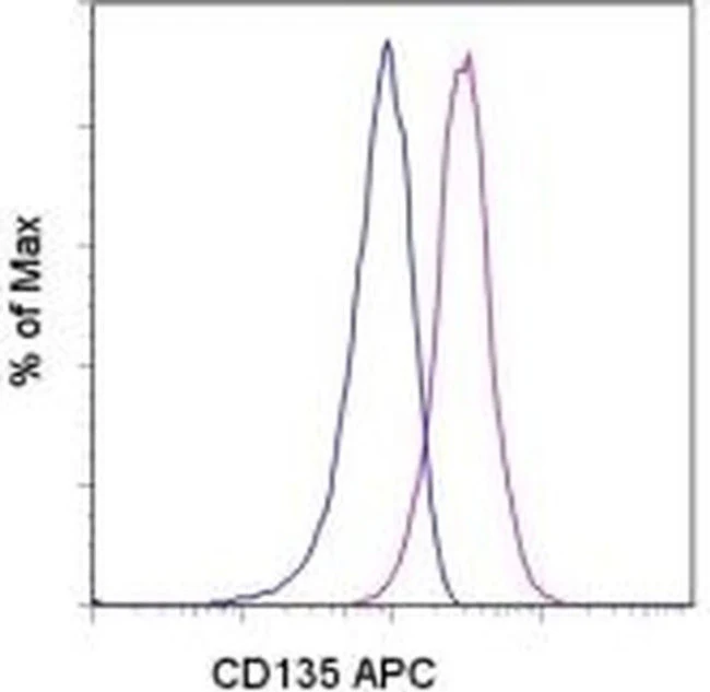
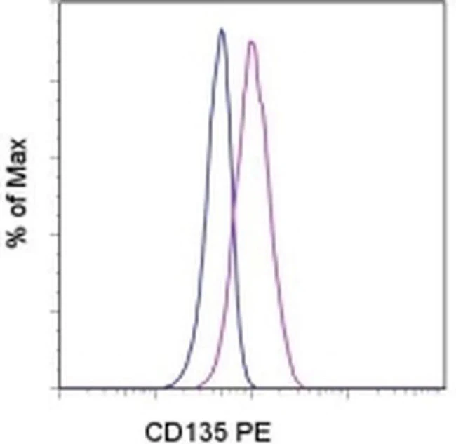
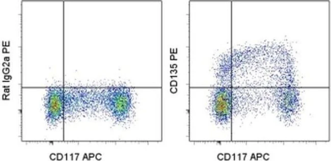
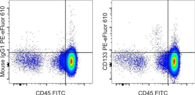
Description: CD115 or Colony-Stimulating Factor-1 Receptor (CSF-1R) is expressed on cells committed to the monocyte lineage and osteoclasts. CSF-1R is a disulfide-linked homodimer, which upon binding of its ligand, CSF-1, undergoes tyrosine autophosphorylation and subsequently, phosphorylates other membrane-proximal downstream targets which results in cytoskeletal remodeling, gene transcription and protein translation. CSF-1R activation promotes the survival, proliferation and differentiation of mononuclear phagocytes and the spreading and motility of macrophages. In osteoclasts, CSF-1R synergizes with RANKL to regulate the differentiation of mononuclear phagocytes to osteoclasts.
Applications Reported: This 12-3A3-1B10 antibody has been reported for use in flow cytometric analysis.
Applications Tested: This 12-3A3-1B10 antibody has been pre-titrated and tested by flow cytometric analysis of normal human peripheral blood cells. This can be used at 5 µL (0.03 µg) per test. A test is defined as the amount (µg) of antibody that will stain a cell sample in a final volume of 100 µL. Cell number should be determined empirically but can range from 10^5 to 10^8 cells/test.
Light sensitivity: This tandem dye is sensitive to photo-induced oxidation. Please protect this vial and stained samples from light.
Fixation: Samples can be stored in IC Fixation Buffer (Product # 00-822-49) (100 µL of cell sample + 100 µL of IC Fixation Buffer) or 1-step Fix/Lyse Solution (Product # 00-5333-54) for up to 3 days in the dark at 4°C with minimal impact on brightness and FRET efficiency/compensation. Some generalizations regarding fluorophore performance after fixation can be made, but clone specific performance should be determined empirically.
Excitation: 488-561 nm; Emission: 775 nm; Laser: Blue Laser, Green Laser, Yellow-Green Laser.
Filtration: 0.2 µm post-manufacturing filtered.
For Research Use Only. Not for use in diagnostic procedures. Not for resale without express authorization.

Description
PRODUCT DETAILS
Host: Rat
Isotype: IgG2a, kappa
Clonality: Monoclonal
Clone: 12-3A3-1B10
Format: PE-Cyanine7
Reactivity: Hu
Application: Flow Cytometry
Tested Dilution: 5 µL (0.03 µg)/test
Concentration: 5 μL/Test
Storage: 4°C, store in dark, DO NOT FREEZE!
Formulation: PBS with BSA and 0.09% sodium azide; pH 7.2
Purification: Affinity chromatography
Data Sheet: TDS
Specific Information
Description: CD115 or Colony-Stimulating Factor-1 Receptor (CSF-1R) is expressed on cells committed to the monocyte lineage and osteoclasts. CSF-1R is a disulfide-linked homodimer, which upon binding of its ligand, CSF-1, undergoes tyrosine autophosphorylation and subsequently, phosphorylates other membrane-proximal downstream targets which results in cytoskeletal remodeling, gene transcription and protein translation. CSF-1R activation promotes the survival, proliferation and differentiation of mononuclear phagocytes and the spreading and motility of macrophages. In osteoclasts, CSF-1R synergizes with RANKL to regulate the differentiation of mononuclear phagocytes to osteoclasts.
Applications Reported: This 12-3A3-1B10 antibody has been reported for use in flow cytometric analysis.
Applications Tested: This 12-3A3-1B10 antibody has been pre-titrated and tested by flow cytometric analysis of normal human peripheral blood cells. This can be used at 5 µL (0.03 µg) per test. A test is defined as the amount (µg) of antibody that will stain a cell sample in a final volume of 100 µL. Cell number should be determined empirically but can range from 10^5 to 10^8 cells/test.
Light sensitivity: This tandem dye is sensitive to photo-induced oxidation. Please protect this vial and stained samples from light.
Fixation: Samples can be stored in IC Fixation Buffer (Product # 00-822-49) (100 µL of cell sample + 100 µL of IC Fixation Buffer) or 1-step Fix/Lyse Solution (Product # 00-5333-54) for up to 3 days in the dark at 4°C with minimal impact on brightness and FRET efficiency/compensation. Some generalizations regarding fluorophore performance after fixation can be made, but clone specific performance should be determined empirically.
Excitation: 488-561 nm; Emission: 775 nm; Laser: Blue Laser, Green Laser, Yellow-Green Laser.
Filtration: 0.2 µm post-manufacturing filtered.
For Research Use Only. Not for use in diagnostic procedures. Not for resale without express authorization.