CD146 Monoclonal Antibody (P1H12), eBioscience
PRODUCT DETAILS
Host: Mouse
Isotype: IgG1, kappa
Clonality: Monoclonal
Clone: P1H12
Reactivity: Ca, Hu, Ms, Rb
Application: Flow Cytometry
Tested Dilution: 0.25 µg/test
Concentration: 0.5 mg/mL
Storage: 4°C
Formulation: PBS with 0.09% sodium azide; pH 7.2
Purification: Affinity chromatography
Data Sheet: TDS
Specific Information
Description: The monoclonal antibody P1H12 recognizes CD146 also known as MUC18, s-endo, Endo-CAM and Mel-CAM, which is a member of the Ig superfamily of proteins. The expression of CD146 is found on endothelial cells, bone marrow fibroblasts and some tumors (especially melanoma). Recently mesenchymal stromal cells and endometrial stromal cells have also been shown to express CD146. The presence of CD146 on circulating blood cells have been confined to a subset of T cells rather than circulating endothelial cells, as expression of other endothelial markers (CD31 and CD51/61) is negative. Expression can be found on activated lymphocytes. The protein is heavily glycosylated with more than 50% of the mass from carbohydrates.
The antibody P1H12 has been reported to crossreact to mouse, rabbit, canine, but not rat.
Applications Reported: This P1H12 antibody has been reported for use in flow cytometric analysis, immunoprecipitation, immunoblotting (WB) under nonreducing conditions, and immunohistology staining of frozen tissue sections.
Applications Tested: This P1H12 antibody has been tested by flow cytometric analysis of HUVEC cell line. This can be used at less than or equal to 0.25 µg per test. A test is defined as the amount (µg) of antibody that will stain a cell sample in a final volume of 100 µL. Cell number should be determined empirically but can range from 10^5 to 10^8 cells/test. It is recommended that the antibody be carefully titrated for optimal performance in the assay of interest.
Purity: Greater than 90%, as determined by SDS-PAGE.
Aggregation: Less than 10%, as determined by HPLC.
Filtration: 0.2 µm post-manufacturing filtered.
For Research Use Only. Not for use in diagnostic procedures. Not for resale without express authorization.
Original: $157.00
-70%$157.00
$47.10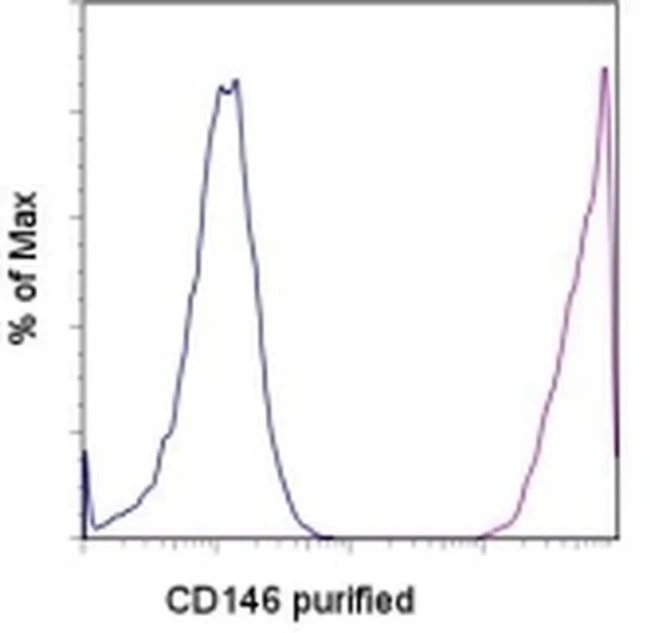
Description
PRODUCT DETAILS
Host: Mouse
Isotype: IgG1, kappa
Clonality: Monoclonal
Clone: P1H12
Reactivity: Ca, Hu, Ms, Rb
Application: Flow Cytometry
Tested Dilution: 0.25 µg/test
Concentration: 0.5 mg/mL
Storage: 4°C
Formulation: PBS with 0.09% sodium azide; pH 7.2
Purification: Affinity chromatography
Data Sheet: TDS
Specific Information
Description: The monoclonal antibody P1H12 recognizes CD146 also known as MUC18, s-endo, Endo-CAM and Mel-CAM, which is a member of the Ig superfamily of proteins. The expression of CD146 is found on endothelial cells, bone marrow fibroblasts and some tumors (especially melanoma). Recently mesenchymal stromal cells and endometrial stromal cells have also been shown to express CD146. The presence of CD146 on circulating blood cells have been confined to a subset of T cells rather than circulating endothelial cells, as expression of other endothelial markers (CD31 and CD51/61) is negative. Expression can be found on activated lymphocytes. The protein is heavily glycosylated with more than 50% of the mass from carbohydrates.
The antibody P1H12 has been reported to crossreact to mouse, rabbit, canine, but not rat.
Applications Reported: This P1H12 antibody has been reported for use in flow cytometric analysis, immunoprecipitation, immunoblotting (WB) under nonreducing conditions, and immunohistology staining of frozen tissue sections.
Applications Tested: This P1H12 antibody has been tested by flow cytometric analysis of HUVEC cell line. This can be used at less than or equal to 0.25 µg per test. A test is defined as the amount (µg) of antibody that will stain a cell sample in a final volume of 100 µL. Cell number should be determined empirically but can range from 10^5 to 10^8 cells/test. It is recommended that the antibody be carefully titrated for optimal performance in the assay of interest.
Purity: Greater than 90%, as determined by SDS-PAGE.
Aggregation: Less than 10%, as determined by HPLC.
Filtration: 0.2 µm post-manufacturing filtered.
For Research Use Only. Not for use in diagnostic procedures. Not for resale without express authorization.