CD279 (PD-1) Monoclonal Antibody (RMP1-30), APC, eBioscience
PRODUCT DETAILS
Host: Rat
Isotype: IgG2b, kappa
Clonality: Monoclonal
Clone: RMP1-30
Format: APC
Reactivity: Ms
Application: Flow Cytometry
Tested Dilution: 1 µg/test
Concentration: 0.2 mg/mL
Storage: 4°C, store in dark, DO NOT FREEZE!
Formulation: PBS with 0.09% sodium azide; pH 7.2
Purification: Affinity chromatography
Data Sheet: TDS
Specific Information
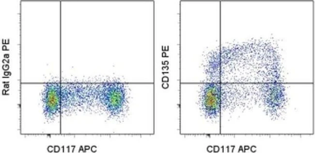
Description: The RMP1-30 antibody reacts with mouse PD-1 (programmed death-1), a 55 kDa member of the Ig superfamily. PD-1 contains the immunoreceptor tyrosine-based inhibitory motif (ITIM) and plays a key role in peripheral tolerance and autoimmune disease in mice. PD-1 is expressed mainly on activated T and B lymphocytes. Two novel B7 Family members have been identified as PD-1 ligands, PD-L1 (B7-H1) and PD-L2 (B7-DC). Evidence reported to date suggests overlapping functions for these ligands and their constitutive expression on some normal tissues and upregulation on activated antigen-presenting cells. RMP1-30 does not block the binding of either B7-H1-Ig or B7-DC-Ig to PD-1 transfectants.
Applications Reported: This RMP1-30 antibody has been reported for use in flow cytometric analysis.
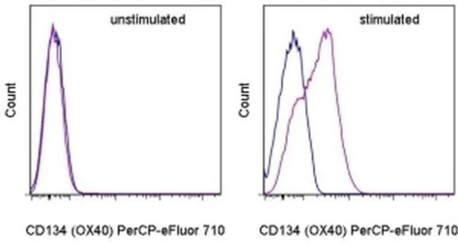
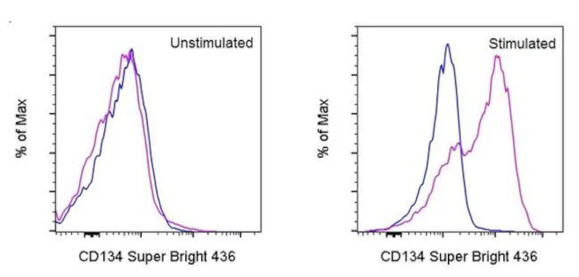
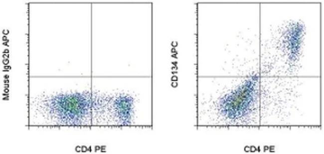
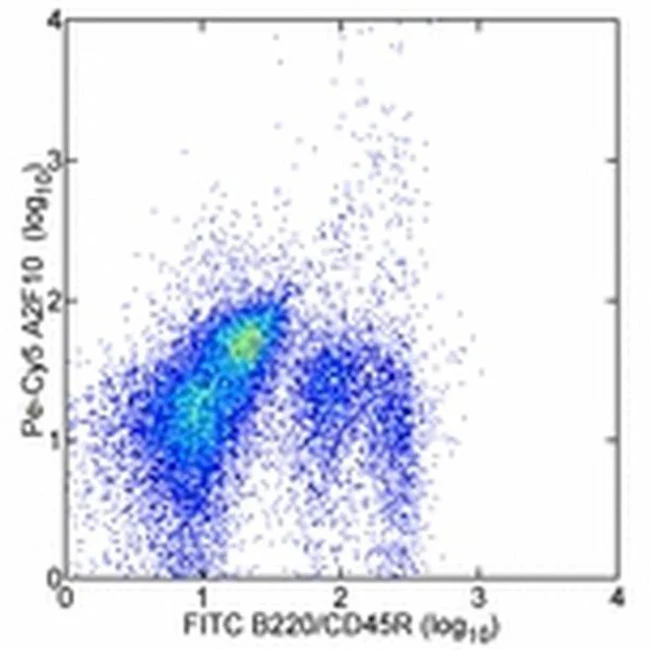
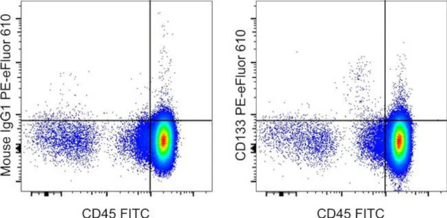
Applications Tested: This RMP1-30 antibody has been tested by flow cytometric analysis of stimulated mouse splenocytes. This can be used at less than or equal to 1 µg per test. A test is defined as the amount (µg) of antibody that will stain a cell sample in a final volume of 100 µL. Cell number should be determined empirically but can range from 10^5 to 10^8 cells/test. It is recommended that the antibody be carefully titrated for optimal performance in the assay of interest.
Excitation: 633-647 nm; Emission: 660 nm; Laser: Red Laser.
Filtration: 0.2 µm post-manufacturing filtered.
For Research Use Only. Not for use in diagnostic procedures. Not for resale without express authorization.

Description
PRODUCT DETAILS
Host: Rat
Isotype: IgG2b, kappa
Clonality: Monoclonal
Clone: RMP1-30
Format: APC
Reactivity: Ms
Application: Flow Cytometry
Tested Dilution: 1 µg/test
Concentration: 0.2 mg/mL
Storage: 4°C, store in dark, DO NOT FREEZE!
Formulation: PBS with 0.09% sodium azide; pH 7.2
Purification: Affinity chromatography
Data Sheet: TDS
Specific Information
Description: The RMP1-30 antibody reacts with mouse PD-1 (programmed death-1), a 55 kDa member of the Ig superfamily. PD-1 contains the immunoreceptor tyrosine-based inhibitory motif (ITIM) and plays a key role in peripheral tolerance and autoimmune disease in mice. PD-1 is expressed mainly on activated T and B lymphocytes. Two novel B7 Family members have been identified as PD-1 ligands, PD-L1 (B7-H1) and PD-L2 (B7-DC). Evidence reported to date suggests overlapping functions for these ligands and their constitutive expression on some normal tissues and upregulation on activated antigen-presenting cells. RMP1-30 does not block the binding of either B7-H1-Ig or B7-DC-Ig to PD-1 transfectants.
Applications Reported: This RMP1-30 antibody has been reported for use in flow cytometric analysis.
Applications Tested: This RMP1-30 antibody has been tested by flow cytometric analysis of stimulated mouse splenocytes. This can be used at less than or equal to 1 µg per test. A test is defined as the amount (µg) of antibody that will stain a cell sample in a final volume of 100 µL. Cell number should be determined empirically but can range from 10^5 to 10^8 cells/test. It is recommended that the antibody be carefully titrated for optimal performance in the assay of interest.
Excitation: 633-647 nm; Emission: 660 nm; Laser: Red Laser.
Filtration: 0.2 µm post-manufacturing filtered.
For Research Use Only. Not for use in diagnostic procedures. Not for resale without express authorization.