CD366 (TIM3) Monoclonal Antibody (RMT3-23), PerCP-eFluor 710, eBioscience
PRODUCT DETAILS
Host: Rat
Isotype: IgG2a, kappa
Clonality: Monoclonal
Clone: RMT3-23
Format: PerCP-eFluor 710
Reactivity: Ms
Application: Flow Cytometry
Tested Dilution: 0.25 µg/test
Concentration: 0.2 mg/mL
Storage: 4°C, store in dark, DO NOT FREEZE!
Formulation: PBS with 0.09% sodium azide; pH 7.2
Purification: Affinity chromatography
Data Sheet: TDS
Specific Information
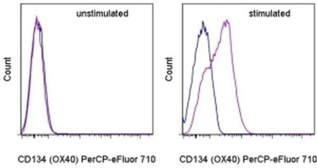
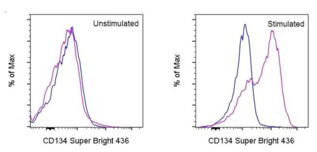
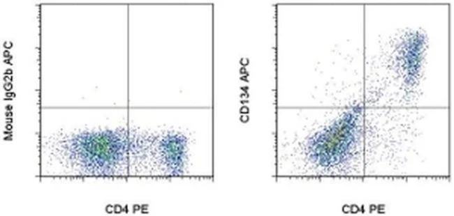
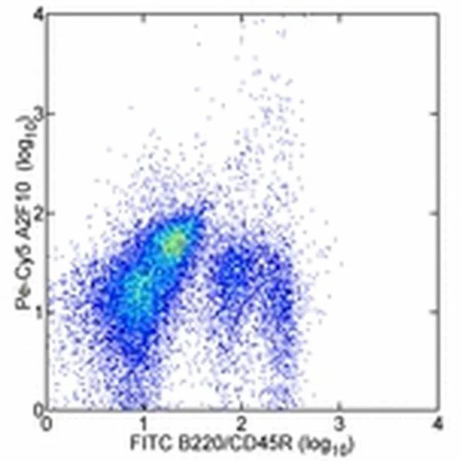
Description: The RMT3-23 monoclonal antibody reacts with mouse CD366 (TIM3), a Th1-specific cell surface protein. The RMT3-23 antibody reacts with CD366 protein expressed by both BALB/c and C57BL/6 strains of mice. CD366, a type I transmembrane protein, contains an immunoglobulin and a mucin-like domain in its extracellular portion and a tyrosine phosphorylation motif in its cytoplasmic portion. CD366 is expressed selectively by differentiated CD4^+Th1 and CD8^+Tc1 cells, but is absent on CD4^+Th2 and CD8^+Tc2 cells. Other hematopoietic cell types, including naive T cells, B cells, macrophages and dendritic cells, do not express CD366, at least at the protein level. CD366 expression is upregulated at a late stage of T cell differentiation on Th1 cells after 3 rounds of in vitro polarization suggesting a role for this molecule in the transport or effector function of Th1 cells rather than a contribution to T cell differentiation. In an experimental autoimmune encephalomyelitis (EAE) model, CD366 was shown to be expressed on most CD4^+ and CD8^+ T cells in the central nervous system at the onset of clinical signs of disease, while less than 2% of CD4^+ cells in the periphery expressed CD366 after immunization.
RMT3-23 has been shown to have functional activity; blocks DC recognition of apoptotic cells and also induces autoantibody production.
Applications Reported: This RMT3-23 antibody has been reported for use in flow cytometric analysis.
Applications Tested: This RMT3-23 antibody has been tested by flow cytometric analysis of mouse splenocytes. This may be used at less than or equal to 0.25 µg per test. A test is defined as the amount (µg) of antibody that will stain a cell sample in a final volume of 100 µL. Cell number should be determined empirically but can range from 10^5 to 10^8 cells/test. It is recommended that the antibody be carefully titrated for optimal performance in the assay of interest.
PerCP-eFluor 710 emits at 710 nm and is excited with the blue laser (488 nm); it can be used in place of PerCP-Cyanine5.5. We recommend using a 710/50 bandpass filter, however, the 695/40 bandpass filter is an acceptable alternative. Please make sure that your instrument is capable of detecting this fluorochrome.
Light sensitivity: This tandem dye is sensitive to photo-induced oxidation. Please protect this vial and stained samples from light.
Fixation: Samples can be stored in IC Fixation Buffer (Product # 00-8222-49) (100 µL of cell sample + 100 µL of IC Fixation Buffer) or 1-step Fix/Lyse Solution (Product # 00-5333-57) for up to 3 days in the dark at 4°C with minimal impact on brightness and FRET efficiency/compensation. Some generalizations regarding fluorophore performance after fixation can be made, but clone specific performance should be determined empirically.
Excitation: 488 nm; Emission: 710 nm; Laser: Blue Laser.
For Research Use Only. Not for use in diagnostic procedures. Not for resale without express authorization.

Description
PRODUCT DETAILS
Host: Rat
Isotype: IgG2a, kappa
Clonality: Monoclonal
Clone: RMT3-23
Format: PerCP-eFluor 710
Reactivity: Ms
Application: Flow Cytometry
Tested Dilution: 0.25 µg/test
Concentration: 0.2 mg/mL
Storage: 4°C, store in dark, DO NOT FREEZE!
Formulation: PBS with 0.09% sodium azide; pH 7.2
Purification: Affinity chromatography
Data Sheet: TDS
Specific Information
Description: The RMT3-23 monoclonal antibody reacts with mouse CD366 (TIM3), a Th1-specific cell surface protein. The RMT3-23 antibody reacts with CD366 protein expressed by both BALB/c and C57BL/6 strains of mice. CD366, a type I transmembrane protein, contains an immunoglobulin and a mucin-like domain in its extracellular portion and a tyrosine phosphorylation motif in its cytoplasmic portion. CD366 is expressed selectively by differentiated CD4^+Th1 and CD8^+Tc1 cells, but is absent on CD4^+Th2 and CD8^+Tc2 cells. Other hematopoietic cell types, including naive T cells, B cells, macrophages and dendritic cells, do not express CD366, at least at the protein level. CD366 expression is upregulated at a late stage of T cell differentiation on Th1 cells after 3 rounds of in vitro polarization suggesting a role for this molecule in the transport or effector function of Th1 cells rather than a contribution to T cell differentiation. In an experimental autoimmune encephalomyelitis (EAE) model, CD366 was shown to be expressed on most CD4^+ and CD8^+ T cells in the central nervous system at the onset of clinical signs of disease, while less than 2% of CD4^+ cells in the periphery expressed CD366 after immunization.
RMT3-23 has been shown to have functional activity; blocks DC recognition of apoptotic cells and also induces autoantibody production.
Applications Reported: This RMT3-23 antibody has been reported for use in flow cytometric analysis.
Applications Tested: This RMT3-23 antibody has been tested by flow cytometric analysis of mouse splenocytes. This may be used at less than or equal to 0.25 µg per test. A test is defined as the amount (µg) of antibody that will stain a cell sample in a final volume of 100 µL. Cell number should be determined empirically but can range from 10^5 to 10^8 cells/test. It is recommended that the antibody be carefully titrated for optimal performance in the assay of interest.
PerCP-eFluor 710 emits at 710 nm and is excited with the blue laser (488 nm); it can be used in place of PerCP-Cyanine5.5. We recommend using a 710/50 bandpass filter, however, the 695/40 bandpass filter is an acceptable alternative. Please make sure that your instrument is capable of detecting this fluorochrome.
Light sensitivity: This tandem dye is sensitive to photo-induced oxidation. Please protect this vial and stained samples from light.
Fixation: Samples can be stored in IC Fixation Buffer (Product # 00-8222-49) (100 µL of cell sample + 100 µL of IC Fixation Buffer) or 1-step Fix/Lyse Solution (Product # 00-5333-57) for up to 3 days in the dark at 4°C with minimal impact on brightness and FRET efficiency/compensation. Some generalizations regarding fluorophore performance after fixation can be made, but clone specific performance should be determined empirically.
Excitation: 488 nm; Emission: 710 nm; Laser: Blue Laser.
For Research Use Only. Not for use in diagnostic procedures. Not for resale without express authorization.