CD63 Monoclonal Antibody (H5C6), eFluor 660, eBioscience
PRODUCT DETAILS
Host: Mouse
Isotype: IgG1, kappa
Clonality: Monoclonal
Clone: H5C6
Format: eFluor 660
Reactivity: Hu
Application: Flow Cytometry
Tested Dilution: 5 µL (0.5 µg)/test
Concentration: 5 μL/Test
Storage: 4°C, store in dark, DO NOT FREEZE!
Formulation: PBS with BSA and 0.09% sodium azide; pH 7.2
Purification: Affinity chromatography
Data Sheet: TDS
Specific Information
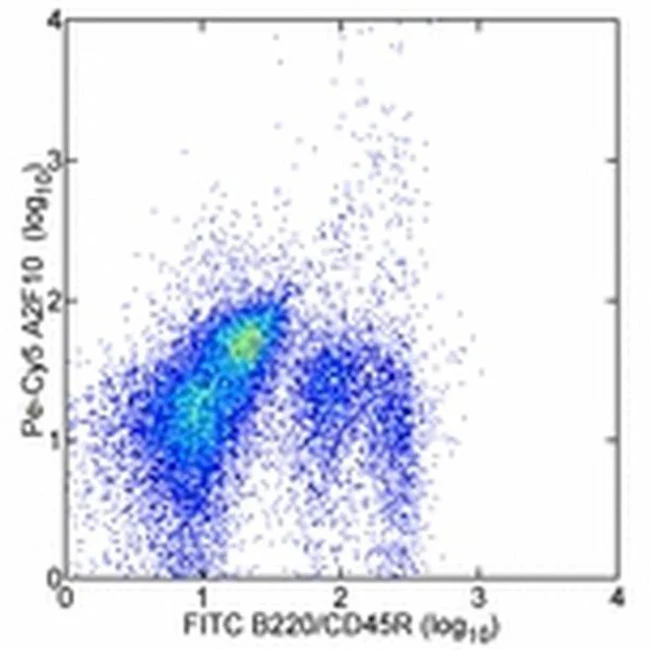
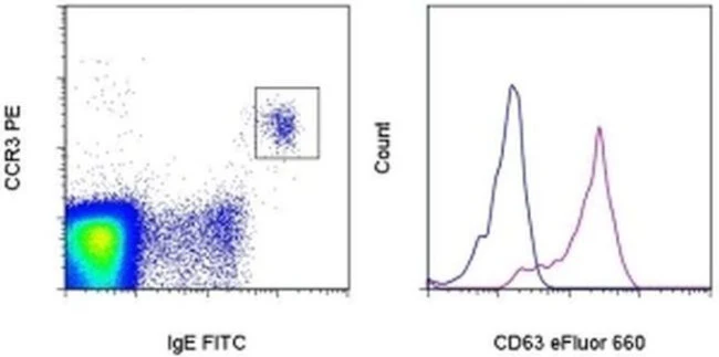
Description: This H5C6 monoclonal antibody reacts with human CD63, a type III member of the tetraspanin family of transmembrane proteins. CD63 is expressed intracellularly on lysosomes, endosomes, and granules of resting platelets and basophils. However, cell surface expression of CD63 can be detected on activated basophils and platelets, monocytes, macrophages, and granulocytes. This receptor is also expressed on endothelial cells, fibroblasts, and smooth muscle cells. Studies have demonstrated that CD63 associates with integrins (VLA-3 and VLA-6) and TIMP-1 to mediate the allergic response.
Applications Reported: This H5C6 antibody has been reported for use in flow cytometric analysis.
Applications Tested: This H5C6 antibody has been pre-titrated and tested by flow cytometric analysis of normal human peripheral blood cells. This can be used at 5 µL (0.5 µg) per test. A test is defined as the amount (µg) of antibody that will stain a cell sample in a final volume of 100 µL. Cell number should be determined empirically but can range from 10^5 to 10^8 cells/test.
eFluor® 660 is a replacement for Alexa Fluor® 647. eFluor® 660 emits at 659 nm and is excited with the red laser (633 nm). Please make sure that your instrument is capable of detecting this fluorochome.
Excitation: 633-647 nm; Emission: 668 nm; Laser: Red Laser.
Filtration: 0.2 µm post-manufacturing filtered.
For Research Use Only. Not for use in diagnostic procedures. Not for resale without express authorization.
Description
PRODUCT DETAILS
Host: Mouse
Isotype: IgG1, kappa
Clonality: Monoclonal
Clone: H5C6
Format: eFluor 660
Reactivity: Hu
Application: Flow Cytometry
Tested Dilution: 5 µL (0.5 µg)/test
Concentration: 5 μL/Test
Storage: 4°C, store in dark, DO NOT FREEZE!
Formulation: PBS with BSA and 0.09% sodium azide; pH 7.2
Purification: Affinity chromatography
Data Sheet: TDS
Specific Information
Description: This H5C6 monoclonal antibody reacts with human CD63, a type III member of the tetraspanin family of transmembrane proteins. CD63 is expressed intracellularly on lysosomes, endosomes, and granules of resting platelets and basophils. However, cell surface expression of CD63 can be detected on activated basophils and platelets, monocytes, macrophages, and granulocytes. This receptor is also expressed on endothelial cells, fibroblasts, and smooth muscle cells. Studies have demonstrated that CD63 associates with integrins (VLA-3 and VLA-6) and TIMP-1 to mediate the allergic response.
Applications Reported: This H5C6 antibody has been reported for use in flow cytometric analysis.
Applications Tested: This H5C6 antibody has been pre-titrated and tested by flow cytometric analysis of normal human peripheral blood cells. This can be used at 5 µL (0.5 µg) per test. A test is defined as the amount (µg) of antibody that will stain a cell sample in a final volume of 100 µL. Cell number should be determined empirically but can range from 10^5 to 10^8 cells/test.
eFluor® 660 is a replacement for Alexa Fluor® 647. eFluor® 660 emits at 659 nm and is excited with the red laser (633 nm). Please make sure that your instrument is capable of detecting this fluorochome.
Excitation: 633-647 nm; Emission: 668 nm; Laser: Red Laser.
Filtration: 0.2 µm post-manufacturing filtered.
For Research Use Only. Not for use in diagnostic procedures. Not for resale without express authorization.