CD63 Monoclonal Antibody (NVG-2), APC, eBioscience
PRODUCT DETAILS
Host: Rat
Isotype: IgG2a, kappa
Clonality: Monoclonal
Clone: NVG-2
Format: APC
Reactivity: Ms
Application: Flow Cytometry
Tested Dilution: 0.25 µg/test
Concentration: 0.2 mg/mL
Storage: 4°C, store in dark, DO NOT FREEZE!
Formulation: PBS with 0.09% sodium azide; pH 7.2
Purification: Affinity chromatography
Data Sheet: TDS
Specific Information
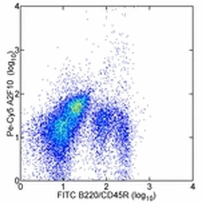
Description: The monoclonal antibody NVG-2 reacts with mouse CD63, also known as Lysosomal-Associated Membrane Protein 3 (LAMP-3) or tetraspanin 30 (TSPN30), a member of tetraspanin family of proteins characterized by four transmembrane domains. CD63 is expressed on a variety of cell types of hematopoietic lineage, e.g., granulocytes, B lymphocytes, platelets, as well as cells of non-hematopoietic origin. It can be found on the cell membrane, late endocytic vesicles, lysosomes, exosomes, and other specialized granules. On the cell surface, CD63 has been shown to interact with various proteins forming tetraspanin-enriched microdomains (TEM). Its high expression on the cell membrane may be indicative of cell activation, hence, CD63 is often used as an activation marker for basophils, platelets and other cells.
Applications Reported: This NVG-2 antibody has been reported for use in intracellular staining followed by flow cytometric analysis.
Applications Tested: This NVG-2 antibody has been tested by intracellular staining followed by flow cytometric analysis of mouse resident peritoneal exudate cells using the Intracellular Fixation & Permeabilization Buffer Set (Product # 88-8824-00) and protocol. Please refer to BestProtocols®: Protocol A: Two step protocol for (cytoplasmic) intracellular proteins located under the Resources Tab online. This can be used at less than or equal to 0.25 µg per test. A test is defined as the amount (µg) of antibody that will stain a cell sample in a final volume of 100 µL. Cell number should be determined empirically but can range from 10^5 to 10^8 cells/test. It is recommended that the antibody be carefully titrated for optimal performance in the assay of interest.
Excitation: 633-647 nm; Emission: 660 nm; Laser: Red Laser.
Filtration: 0.2 µm post-manufacturing filtered.
For Research Use Only. Not for use in diagnostic procedures. Not for resale without express authorization.

Description
PRODUCT DETAILS
Host: Rat
Isotype: IgG2a, kappa
Clonality: Monoclonal
Clone: NVG-2
Format: APC
Reactivity: Ms
Application: Flow Cytometry
Tested Dilution: 0.25 µg/test
Concentration: 0.2 mg/mL
Storage: 4°C, store in dark, DO NOT FREEZE!
Formulation: PBS with 0.09% sodium azide; pH 7.2
Purification: Affinity chromatography
Data Sheet: TDS
Specific Information
Description: The monoclonal antibody NVG-2 reacts with mouse CD63, also known as Lysosomal-Associated Membrane Protein 3 (LAMP-3) or tetraspanin 30 (TSPN30), a member of tetraspanin family of proteins characterized by four transmembrane domains. CD63 is expressed on a variety of cell types of hematopoietic lineage, e.g., granulocytes, B lymphocytes, platelets, as well as cells of non-hematopoietic origin. It can be found on the cell membrane, late endocytic vesicles, lysosomes, exosomes, and other specialized granules. On the cell surface, CD63 has been shown to interact with various proteins forming tetraspanin-enriched microdomains (TEM). Its high expression on the cell membrane may be indicative of cell activation, hence, CD63 is often used as an activation marker for basophils, platelets and other cells.
Applications Reported: This NVG-2 antibody has been reported for use in intracellular staining followed by flow cytometric analysis.
Applications Tested: This NVG-2 antibody has been tested by intracellular staining followed by flow cytometric analysis of mouse resident peritoneal exudate cells using the Intracellular Fixation & Permeabilization Buffer Set (Product # 88-8824-00) and protocol. Please refer to BestProtocols®: Protocol A: Two step protocol for (cytoplasmic) intracellular proteins located under the Resources Tab online. This can be used at less than or equal to 0.25 µg per test. A test is defined as the amount (µg) of antibody that will stain a cell sample in a final volume of 100 µL. Cell number should be determined empirically but can range from 10^5 to 10^8 cells/test. It is recommended that the antibody be carefully titrated for optimal performance in the assay of interest.
Excitation: 633-647 nm; Emission: 660 nm; Laser: Red Laser.
Filtration: 0.2 µm post-manufacturing filtered.
For Research Use Only. Not for use in diagnostic procedures. Not for resale without express authorization.