CD68 Monoclonal Antibody (FA-11), PE, eBioscience
PRODUCT DETAILS
Host: Rat
Isotype: IgG2a, kappa
Clonality: Monoclonal
Clone: FA-11
Format: PE
Reactivity: Ms
Application: Flow Cytometry
Tested Dilution: 0.25 µg/test
Concentration: 0.2 mg/mL
Storage: 4°C, store in dark, DO NOT FREEZE!
Formulation: PBS with 0.09% sodium azide; pH 7.2
Purification: Affinity chromatography
Data Sheet: TDS
Specific Information
Description: This FA-11 monoclonal antibody reacts with mouse CD68, known also as macrosialin. CD68 is a heavily O- and N- glycosylated type I integral membrane protein belonging to the sialomucin family and is closely related to the Lysosomal/endosomal-Associated Membrane Glycoproteins (LAMP). Although commonly used as a pan-macrophage marker, low levels of expression have also been observed in Langerhans cells and conventional dendritic cells. CD68 is mostly localized in the late endosomes and lysosomes with a smaller fraction circulating to the cell surface. In the steady state, only about 10-15% of CD68 is present at the surface. Cell stimulation increases expression of CD68, its transport to cell surface, and results in glycoform remodeling. CD68 binds oxidized LDL and is involved in the process of phagocytosis.
Applications Reported: This FA-11 antibody has been reported for use in intracellular staining followed by flow cytometric analysis.
Applications Tested: This FA-11 antibody has been tested by intracellular staining and flow cytometric analysis of mouse bone marrow derived macrophages using the Intracellular Fixation & Permeabilization Buffer Set (Product # 88-8824-00) and protocol. Please refer to BestProtocols®: Protocol A: Two step protocol for (cytoplasmic) intracellular proteins located under the Resources Tab online. This can be used at less than or equal to 0.25 µg per test. A test is defined as the amount (µg) of antibody that will stain a cell sample in a final volume of 100 µL. Cell number should be determined empirically but can range from 10^5 to 10^8 cells/test. It is recommended that the antibody be carefully titrated for optimal performance in the assay of interest.
Excitation: 488-561 nm; Emission: 578 nm; Laser: Blue Laser, Green Laser, Yellow-Green Laser.
Filtration: 0.2 µm post-manufacturing filtered.
For Research Use Only. Not for use in diagnostic procedures. Not for resale without express authorization.
Original: $411.00
-70%$411.00
$123.30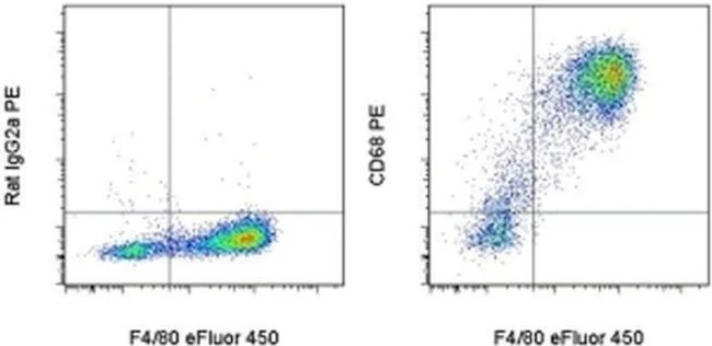
Description
PRODUCT DETAILS
Host: Rat
Isotype: IgG2a, kappa
Clonality: Monoclonal
Clone: FA-11
Format: PE
Reactivity: Ms
Application: Flow Cytometry
Tested Dilution: 0.25 µg/test
Concentration: 0.2 mg/mL
Storage: 4°C, store in dark, DO NOT FREEZE!
Formulation: PBS with 0.09% sodium azide; pH 7.2
Purification: Affinity chromatography
Data Sheet: TDS
Specific Information
Description: This FA-11 monoclonal antibody reacts with mouse CD68, known also as macrosialin. CD68 is a heavily O- and N- glycosylated type I integral membrane protein belonging to the sialomucin family and is closely related to the Lysosomal/endosomal-Associated Membrane Glycoproteins (LAMP). Although commonly used as a pan-macrophage marker, low levels of expression have also been observed in Langerhans cells and conventional dendritic cells. CD68 is mostly localized in the late endosomes and lysosomes with a smaller fraction circulating to the cell surface. In the steady state, only about 10-15% of CD68 is present at the surface. Cell stimulation increases expression of CD68, its transport to cell surface, and results in glycoform remodeling. CD68 binds oxidized LDL and is involved in the process of phagocytosis.
Applications Reported: This FA-11 antibody has been reported for use in intracellular staining followed by flow cytometric analysis.
Applications Tested: This FA-11 antibody has been tested by intracellular staining and flow cytometric analysis of mouse bone marrow derived macrophages using the Intracellular Fixation & Permeabilization Buffer Set (Product # 88-8824-00) and protocol. Please refer to BestProtocols®: Protocol A: Two step protocol for (cytoplasmic) intracellular proteins located under the Resources Tab online. This can be used at less than or equal to 0.25 µg per test. A test is defined as the amount (µg) of antibody that will stain a cell sample in a final volume of 100 µL. Cell number should be determined empirically but can range from 10^5 to 10^8 cells/test. It is recommended that the antibody be carefully titrated for optimal performance in the assay of interest.
Excitation: 488-561 nm; Emission: 578 nm; Laser: Blue Laser, Green Laser, Yellow-Green Laser.
Filtration: 0.2 µm post-manufacturing filtered.
For Research Use Only. Not for use in diagnostic procedures. Not for resale without express authorization.