CD68 Monoclonal Antibody (FA-11), PerCP-eFluor 710, eBioscience
PRODUCT DETAILS
Host: Rat
Isotype: IgG2a, kappa
Clonality: Monoclonal
Clone: FA-11
Format: PerCP-eFluor 710
Reactivity: Ms
Application: Flow Cytometry
Tested Dilution: 0.25 µg/test
Concentration: 0.2 mg/mL
Storage: 4°C, store in dark, DO NOT FREEZE!
Formulation: PBS with 0.09% sodium azide; pH 7.2
Purification: Affinity chromatography
Data Sheet: TDS
Specific Information
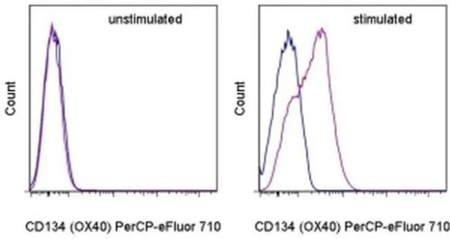
Description: This FA-11 monoclonal antibody reacts with mouse CD68, known also as macrosialin. CD68 is a heavily O- and N- glycosylated type I integral membrane protein belonging to the sialomucin family and is closely related to the Lysosomal/endosomal-Associated Membrane Glycoproteins (LAMP). Although commonly used as a pan-macrophage marker, low levels of expression have also been observed in Langerhans cells and conventional dendritic cells. CD68 is mostly localized in the late endosomes and lysosomes with a smaller fraction circulating to the cell surface. In the steady state, only about 10-15% of CD68 is present at the surface. Cell stimulation increases expression of CD68, its transport to cell surface, and results in glycoform remodeling. CD68 binds oxidized LDL and is involved in the process of phagocytosis.
Applications Reported: This FA-11 antibody has been reported for use in intracellular staining followed by flow cytometric analysis.
Applications Tested: This FA-11 antibody has been tested by intracellular staining followed by flow cytometric analysis of mouse bone marrow cells using the Intracellular Fixation & Permeabilization Buffer Set (Product # 88-8824-00) and protocol. Please refer to "Staining Intracellular Antigens for Flow Cytometry, Protocol A: Two step protocol for intracellular (cytoplasmic) proteins" located at www.thermofisher.com/flowprotocols . This may be used at less than or equal to 0.25 µg per test. A test is defined as the amount (µg) of antibody that will stain a cell sample in a final volume of 100 µL. Cell number should be determined empirically but can range from 10^5 to 10^8 cells/test. It is recommended that the antibody be carefully titrated for optimal performance in the assay of interest.
PerCP-eFluor 710 emits at 710 nm and is excited with the blue laser (488 nm); it can be used in place of PerCP-Cyanine5.5. We recommend using a 710/50 bandpass filter, however, the 695/40 bandpass filter is an acceptable alternative. Please make sure that your instrument is capable of detecting this fluorochrome.
Light sensitivity: This tandem dye is sensitive to photo-induced oxidation. Please protect this vial and stained samples from light.
Fixation: Samples can be stored in IC Fixation Buffer (Product # 00-8222-49) (100 µL of cell sample + 100 µL of IC Fixation Buffer) or 1-step Fix/Lyse Solution (Product # 00-5333-57) for up to 3 days in the dark at 4°C with minimal impact on brightness and FRET efficiency/compensation. Some generalizations regarding fluorophore performance after fixation can be made, but clone specific performance should be determined empirically.
Excitation: 488 nm; Emission: 710 nm; Laser: Blue Laser.
For Research Use Only. Not for use in diagnostic procedures. Not for resale without express authorization.
Original: $436.00
-70%$436.00
$130.80
Description
PRODUCT DETAILS
Host: Rat
Isotype: IgG2a, kappa
Clonality: Monoclonal
Clone: FA-11
Format: PerCP-eFluor 710
Reactivity: Ms
Application: Flow Cytometry
Tested Dilution: 0.25 µg/test
Concentration: 0.2 mg/mL
Storage: 4°C, store in dark, DO NOT FREEZE!
Formulation: PBS with 0.09% sodium azide; pH 7.2
Purification: Affinity chromatography
Data Sheet: TDS
Specific Information
Description: This FA-11 monoclonal antibody reacts with mouse CD68, known also as macrosialin. CD68 is a heavily O- and N- glycosylated type I integral membrane protein belonging to the sialomucin family and is closely related to the Lysosomal/endosomal-Associated Membrane Glycoproteins (LAMP). Although commonly used as a pan-macrophage marker, low levels of expression have also been observed in Langerhans cells and conventional dendritic cells. CD68 is mostly localized in the late endosomes and lysosomes with a smaller fraction circulating to the cell surface. In the steady state, only about 10-15% of CD68 is present at the surface. Cell stimulation increases expression of CD68, its transport to cell surface, and results in glycoform remodeling. CD68 binds oxidized LDL and is involved in the process of phagocytosis.
Applications Reported: This FA-11 antibody has been reported for use in intracellular staining followed by flow cytometric analysis.
Applications Tested: This FA-11 antibody has been tested by intracellular staining followed by flow cytometric analysis of mouse bone marrow cells using the Intracellular Fixation & Permeabilization Buffer Set (Product # 88-8824-00) and protocol. Please refer to "Staining Intracellular Antigens for Flow Cytometry, Protocol A: Two step protocol for intracellular (cytoplasmic) proteins" located at www.thermofisher.com/flowprotocols . This may be used at less than or equal to 0.25 µg per test. A test is defined as the amount (µg) of antibody that will stain a cell sample in a final volume of 100 µL. Cell number should be determined empirically but can range from 10^5 to 10^8 cells/test. It is recommended that the antibody be carefully titrated for optimal performance in the assay of interest.
PerCP-eFluor 710 emits at 710 nm and is excited with the blue laser (488 nm); it can be used in place of PerCP-Cyanine5.5. We recommend using a 710/50 bandpass filter, however, the 695/40 bandpass filter is an acceptable alternative. Please make sure that your instrument is capable of detecting this fluorochrome.
Light sensitivity: This tandem dye is sensitive to photo-induced oxidation. Please protect this vial and stained samples from light.
Fixation: Samples can be stored in IC Fixation Buffer (Product # 00-8222-49) (100 µL of cell sample + 100 µL of IC Fixation Buffer) or 1-step Fix/Lyse Solution (Product # 00-5333-57) for up to 3 days in the dark at 4°C with minimal impact on brightness and FRET efficiency/compensation. Some generalizations regarding fluorophore performance after fixation can be made, but clone specific performance should be determined empirically.
Excitation: 488 nm; Emission: 710 nm; Laser: Blue Laser.
For Research Use Only. Not for use in diagnostic procedures. Not for resale without express authorization.