Dendritic Cell Marker DCIR2 Monoclonal Antibody (33D1), eBioscience
PRODUCT DETAILS
Host: Rat
Isotype: IgG2b, kappa
Clonality: Monoclonal
Clone: 33D1
Reactivity: Ms
Application: Flow Cytometry
Tested Dilution: 1 µg/test
Concentration: 0.5 mg/mL
Storage: 4°C
Formulation: PBS with 0.09% sodium azide; pH 7.2
Purification: Affinity chromatography
Data Sheet: TDS
Specific Information
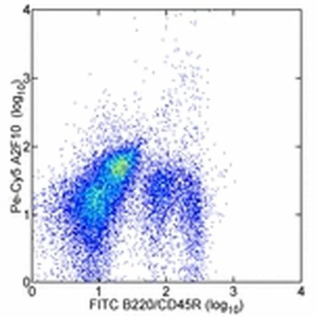
Description: The 33D1 monoclonal antibody reacts with a mouse dendritic cell (DC)-specific surface marker, DCIR2. The nature and biological activity of DCIR2 is unknown. DCIR2 has been reported on a variety of dendritic cell subpopulations from mouse thymus, spleen, lymph node, and Peyer's patch. Bone marrow dendritic cells require GM-CSF to express DCIR2, and this expression is downregulated in the presence of IL-4. DCIR2 has been detected in vivo in brain dendritic cells post infection with Toxoplasma gondii.
Applications Reported: This 33D1 antibody has been reported for use in flow cytometric analysis, immunohistology staining of frozen tissue sections, and immunohistochemical staining. It has also been reported in complement-mediated cytotoxicity.
Applications Tested: The 33D1 antibody has been tested by flow cytometric analysis of mouse splenocytes. This can be used at less than or equal to 1 µg per test. A test is defined as the amount (µg) of antibody that will stain a cell sample in a final volume of 100 µL. Cell number should be determined empirically but can range from 10^5 to 10^8 cells/test. It is recommended that the antibody be carefully titrated for optimal performance in the assay of interest. For detection of purified 33D1 it is recommended not to use goat anti-rat due to its low affinity for 33D1. Rather, we recommend using biotin mouse anti-rat (Product # 13-4813-85).
Purity: Greater than 90%, as determined by SDS-PAGE.
Aggregation: Less than 10%, as determined by HPLC.
Filtration: 0.2 µm post-manufacturing filtered.
For Research Use Only. Not for use in diagnostic procedures. Not for resale without express authorization.
Original: $128.00
-70%$128.00
$38.40
Description
PRODUCT DETAILS
Host: Rat
Isotype: IgG2b, kappa
Clonality: Monoclonal
Clone: 33D1
Reactivity: Ms
Application: Flow Cytometry
Tested Dilution: 1 µg/test
Concentration: 0.5 mg/mL
Storage: 4°C
Formulation: PBS with 0.09% sodium azide; pH 7.2
Purification: Affinity chromatography
Data Sheet: TDS
Specific Information
Description: The 33D1 monoclonal antibody reacts with a mouse dendritic cell (DC)-specific surface marker, DCIR2. The nature and biological activity of DCIR2 is unknown. DCIR2 has been reported on a variety of dendritic cell subpopulations from mouse thymus, spleen, lymph node, and Peyer's patch. Bone marrow dendritic cells require GM-CSF to express DCIR2, and this expression is downregulated in the presence of IL-4. DCIR2 has been detected in vivo in brain dendritic cells post infection with Toxoplasma gondii.
Applications Reported: This 33D1 antibody has been reported for use in flow cytometric analysis, immunohistology staining of frozen tissue sections, and immunohistochemical staining. It has also been reported in complement-mediated cytotoxicity.
Applications Tested: The 33D1 antibody has been tested by flow cytometric analysis of mouse splenocytes. This can be used at less than or equal to 1 µg per test. A test is defined as the amount (µg) of antibody that will stain a cell sample in a final volume of 100 µL. Cell number should be determined empirically but can range from 10^5 to 10^8 cells/test. It is recommended that the antibody be carefully titrated for optimal performance in the assay of interest. For detection of purified 33D1 it is recommended not to use goat anti-rat due to its low affinity for 33D1. Rather, we recommend using biotin mouse anti-rat (Product # 13-4813-85).
Purity: Greater than 90%, as determined by SDS-PAGE.
Aggregation: Less than 10%, as determined by HPLC.
Filtration: 0.2 µm post-manufacturing filtered.
For Research Use Only. Not for use in diagnostic procedures. Not for resale without express authorization.